Dental Implant Planning: Why the Final Tooth Comes First
A dental implant supports the replacement tooth that the patient will ultimately see, use, and maintain. That restoration may be a single implant crown, an implant-supported bridge, an overdenture, or a full-arch restoration.
For that reason, dental implant planning should not begin with only one question: Where can an implant fit?
It should also ask: Where does the final tooth need to be?
Once the intended tooth position is established, the available bone, gum tissue, bite, neighboring teeth, and important anatomical structures can be evaluated in relation to that goal.
Successful integration with the bone is essential. However, integration alone does not guarantee a successful restorative result. The implant must also support a restoration with appropriate function, esthetics, contours, and access for cleaning.
Implant position and angulation can influence:
• The shape and size of the implant crown
• How the crown meets the gumline
• The location of the screw-access opening
• The relationship with neighboring and opposing teeth
• How forces are distributed through the bite
• The patient’s ability to clean around the restoration
An implant may integrate successfully and still create restorative limitations if its position does not support the planned tooth. The final crown may require a bulky or difficult-to-clean contour, the screw-access opening may emerge in an undesirable location, or the bite may become more difficult to manage.
Digital implant planning allows the intended restoration to be studied in relation to the patient’s anatomy before surgery.
A CBCT scan, which is a three-dimensional dental scan, helps evaluate bone dimensions and important anatomical boundaries at the proposed implant site. An intraoral scan records the teeth, surface contours, and bite. A diagnostic tooth design, sometimes called a digital wax-up, shows the intended position, shape, and size of the replacement tooth.
When these records are combined, I can evaluate the proposed implant position in relation to both the planned restoration and the available anatomy.
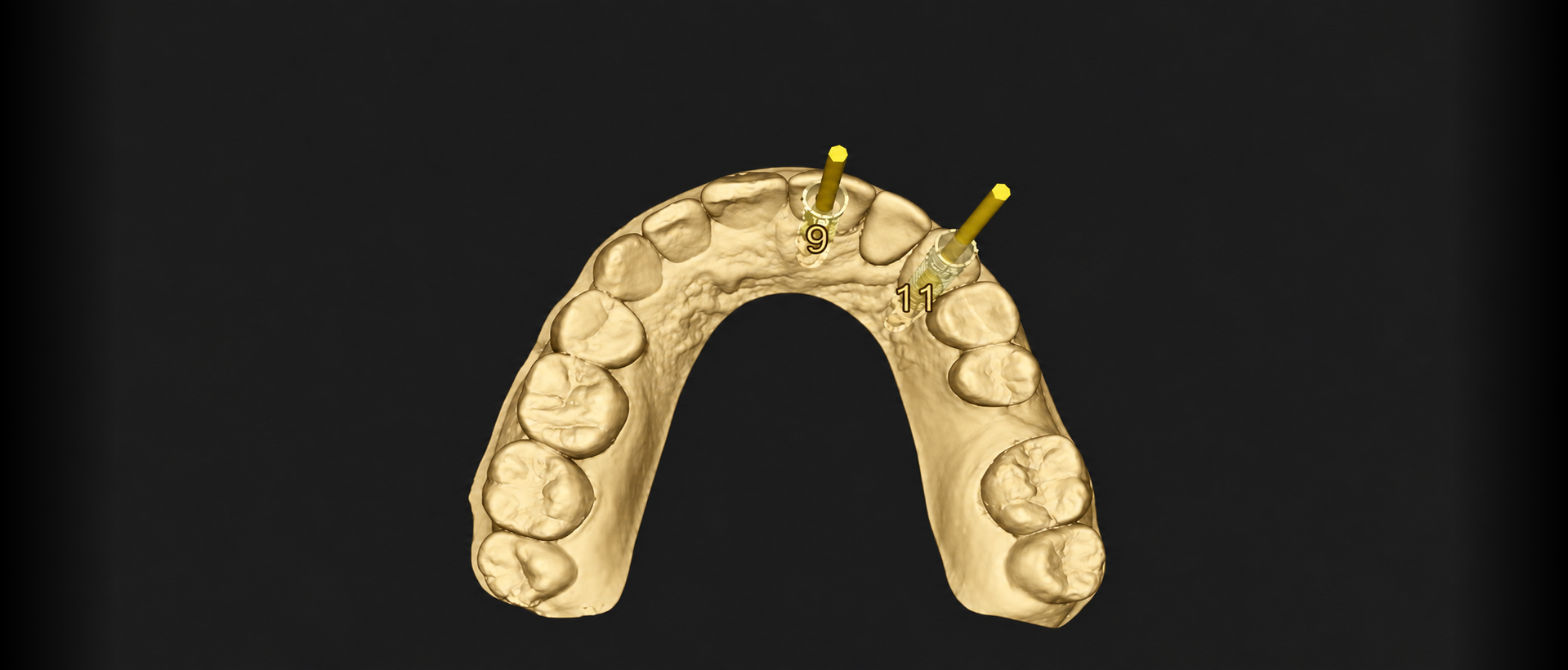
The accompanying image illustrates how proposed implant positions can be reviewed in relation to the maxillary arch and intended restorations before treatment begins.
Digital records provide valuable information, but they do not replace the clinical examination or professional judgment.
The planning requirements differ depending on the location and type of restoration.
For a front tooth in the esthetic zone, dental implant planning may include how much tooth and gum tissue show when the patient smiles, lip position, gum levels, tissue thickness, neighboring teeth, restorative contours, and the opposing bite.
For a back tooth, the plan must account for chewing surfaces, available restorative space, adjacent roots, opposing teeth, and the direction of functional forces.
The intended tooth position must then be reconciled with the patient’s anatomy. Bone dimensions, site characteristics, sinus position, nerves, neighboring roots, and soft-tissue conditions can all influence what is possible.
Sometimes the planned restoration must be modified. In other cases, bone or soft-tissue grafting may be considered. Occasionally, an implant may not be the most appropriate treatment.
The same principles become even more important when several implants will support a bridge or full-arch restoration. The planned tooth arrangement, restorative space, hygiene access, and distribution of forces must be evaluated across the arch rather than one implant at a time.
Careful planning helps identify these issues before surgery rather than after an implant has already been placed.
When appropriate, a surgical guide can help translate a digital implant plan to the surgical site.
The guide is designed using the combined anatomical and restorative records. It can assist with transferring the planned implant position, direction, and depth during guided implant surgery. Some deviation from the digital plan can still occur.
A surgical guide is not necessary for every case, and it does not replace the surgeon’s clinical judgment. The bone, gum tissue, implant stability, and conditions encountered during surgery must still be evaluated.
When another specialist performs the surgery, I prefer that collaboration to begin during planning. The restorative requirements can then be coordinated among the prosthodontist, surgeon, and dental laboratory before the implant position is finalized.
An implant restoration must do more than look appropriate in a digital design.
The opposing teeth, available restorative space, chewing pattern, functional movements, speech, and overall bite may influence the final design. The restoration must also allow reasonable access for daily cleaning and professional maintenance.
For more complex cases, a provisional, or temporary, restoration may provide useful clinical information before the final prosthesis is completed. It can help evaluate appearance, speech, comfort, tissue response, function, and the way the teeth come together.
In selected cases, the provisional restoration is part of the clinical evaluation rather than simply a temporary cosmetic replacement.
As a prosthodontist, I plan the replacement tooth and evaluate how the implant, restoration, bite, gum tissue, and long-term maintenance need to work together.
At Smile Boutique Implant Center in Austin, I use this restoration-driven approach to coordinate the prosthodontic and surgical phases from the beginning.
The surgery serves the restorative plan. The final restoration should not have to compensate for a decision that could have been addressed before surgery.
This article is educational and is not a substitute for an individual examination, diagnosis, or treatment recommendation.